Ziekteleer spijsvertering
Praktisch
- 6 stp
- eerste semester
- maag-darm
- lever-galblaas-pancreas
-
radiologie
- prof. L. Konijn
- examenvragen
-
endoscopie
- prof. I. Demedts
- examenvragen
- examen
- verdeling examenvragen in oude mastervak
- 11/50 Laleman - lever
- 10/50 D'Hoore - heelkunde
- 8/50 De Hertogh + Roskams - pathologie
- 7/50 Konijn - radiologie
- 6/50 Topal - onco
- 6/50 Tack - maag/darm
- 2/50 urgentieweek (n.v.t)
- 0/50 Demedts - endoscopie
- 0/50 Ferrante - IBD
- nieuwe vak
-
prof. Laleman (11/50)
- elk jaar nieuwe vragen
- voorbeelden + weggevers: laatste les
- beste leverfuncietest: PT (of INR)
- welke hepatitis nooit chronisch: hepatitis A
- E normaal ook niet, maar wel bij ...
-
casus virale hepatitis + serologie
kenmerk hepatitis A hepatitis B hepatitis C hepatitis E transmissie • fecaal-oraal
• direct contact• lichaamsvochten
• SOA
• besmet bloed
• injecties
• verticaalidem HBV idem HAV incubatieduur 2–6 w 2–6 m 2 w–6 m idem HAV chroniciteit nee • 90% bij primo-infectie als kind
• 2–6% als volwassene50% nee, tenzij immuungecompromitteerd vaccin • Havrix: 0 en 6 m
• Twinrix: 0, 1 en 6 m• Engerix: 0, 1 en 6 m
• Twinrix: 0, 1 en 6 m- - virus acuut chronisch HAV supportief: lichte maaltijden, hygiëne; geen antivirale therapie - HBV opvolgen; antiviraal bij PT < 50% en fulminante/icterische hepatitis antiviraal bij hepatitisfase: Entecavir / Tenofovir HCV zelden tot nooit behandelen altijd direct antiviral agents (DAAs) HEV cf. HAV: supportief - afkorting betekenis HBsAg vroegste marker van infectie; bij > 6 m positief: chronische infectie HBsAb marker van immuniteit; levenslang bij > 10 IU/mL; verschijnt na 1–4 m HBcAb marker van huidige of vroegere blootstelling aan HBV HBcAb-IgM marker van recente blootstelling aan HBV HBeAg marker van actieve replicatie; lager bij patiënten met een precore-mutatie HBeAb seroconversie vanuit HBeAg-positiviteit; wijst relatief op herstel en verminderde besmettelijkheid HBV-DNA index voor actieve virale replicatie scenario HBsAg anti-HBs IgM-anti-HBc IgG-anti-HBc HBeAg anti-HBe HBV-DNA ALT HBV-naïef 0 0 0 0 0 0 0 normaal HBV-vaccinatie (immuniteit) 0 + 0 0 0 0 0 normaal acute hepatitis B + 0 + −/+ + 0 +++ verhoogd herstel van hepatitis B (immuniteit) 0 + 0 + 0 + 0 normaal chronische (eAg+/eAg−) hepatitis B-infectie + > 6 m 0 0 + +/− −/+ +++/+ normaal chronische (eAg+/eAg−) hepatitis B + > 6 m 0 0 + +/− −/+ +++/++ verhoogd
-
casus ascites
totaal eiwit ascites SAAG ≥ 1,1 g/dL — portale hypertensie SAAG < 1,1 g/dL < 2,5 g/dL prehepatisch/hepatisch:
• cirrose
• infiltratieve ziekten
• v. portae-trombose
• schistosomiasis• nefrotisch syndroom
• protein-losing enteropathy≥ 2,5 g/dL posthepatisch:
• Budd-Chiari, vroege fase
• hartfalen
• pericarditis constrictiva• peritoneale carcinomatose
• pancreatische ascites
• peritoneale tuberculose
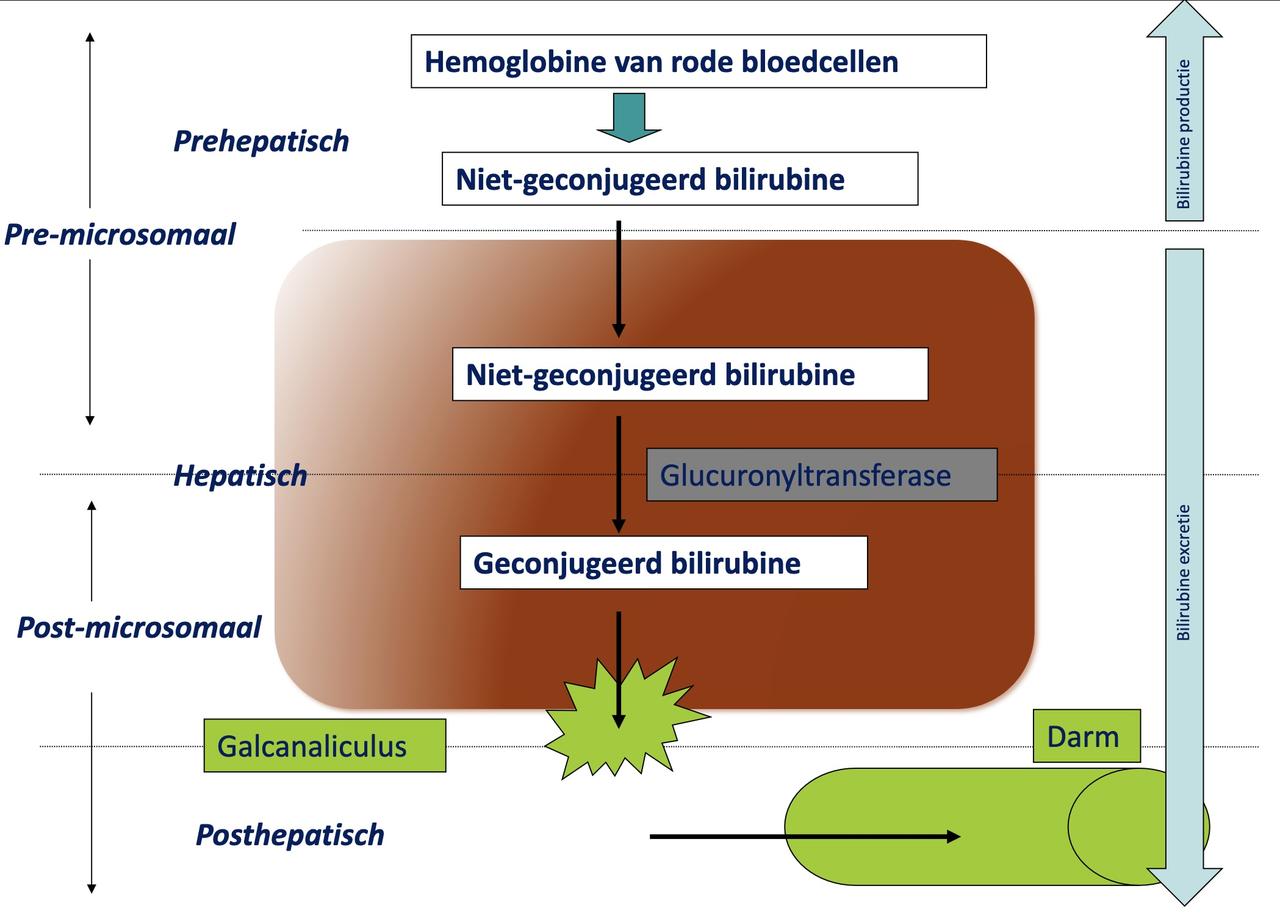
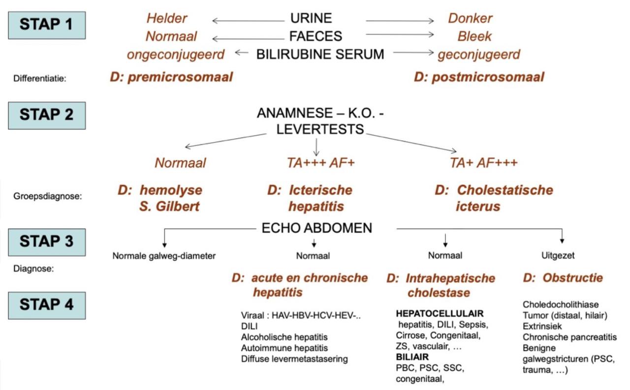
- casus icterus
-
- prof. Konijn
- 7/50 vragen
- 3 theorie
- 4 vragen met plaatjes
- prof. De Hertogh
- wel histologische beelden
- veel hergebruik oude examenvragen - prof. Roskams
- geen histologische beelden
- kenmerken van cholestatische ziektes, hepatitis of tumoren als MCQ - prof. Tack
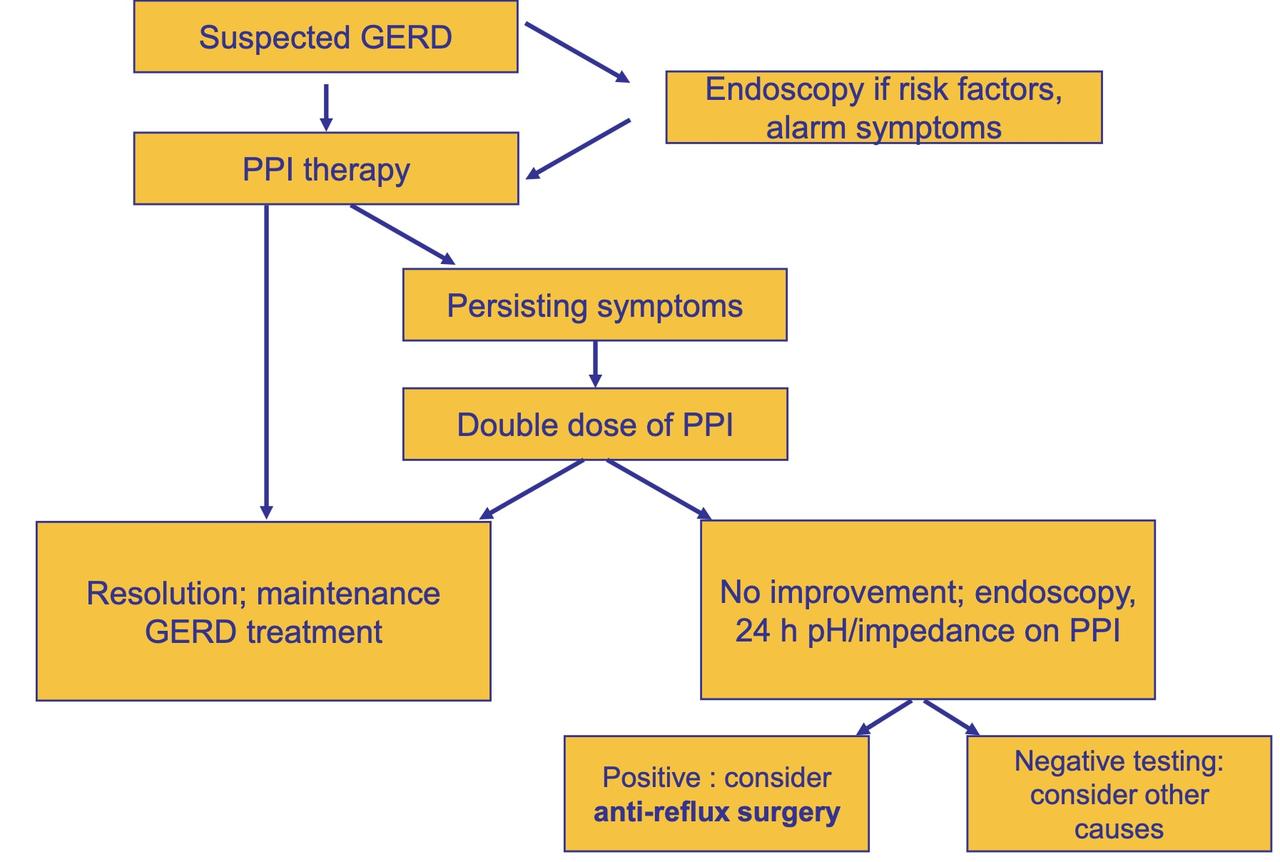
- GERD
- risicofactoren
- 50j+
- roken
- alcohol
- NSAID
- familiale voorgeschiedenis
- alarmsignalen
- dysfagie
- vermagering
- bloedverlies
- (nachtelijke klachten)
- risicofactoren
-
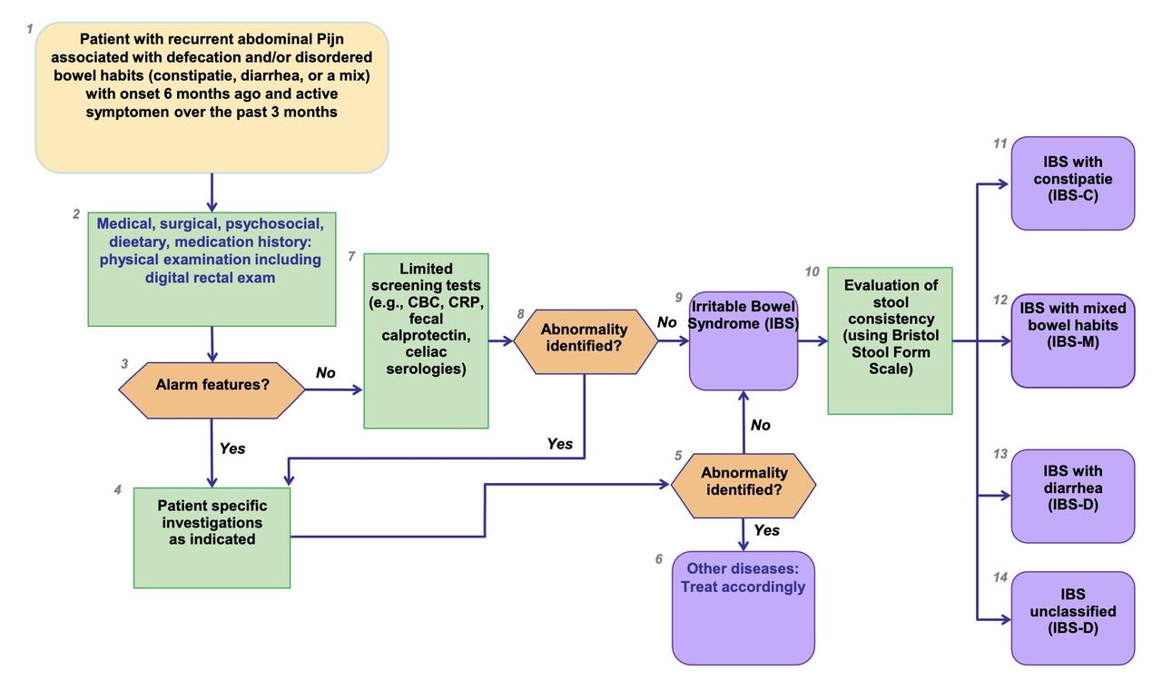
- alarmtekens
- anaal bloedverlies
- vermagering
- koorts
- anemia
- familiaal: darmkanker, IBD, coeliakie
- afwijkend KO
- alarmtekens
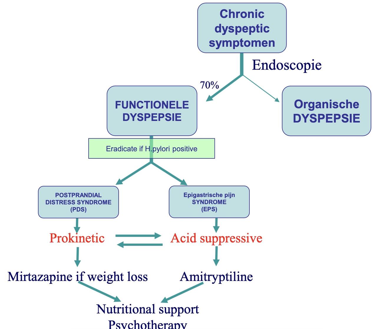
- wikimedica
- oud curriculum (1e master, 8stp)
- nieuw curriculum
- reco januari 2026
- TODO
- prof. Tack: klinische lessen
- prof. Topal: SMD tumoren
- prof. De Hertogh: les 3/3
- prof. Konijn
- samenvatting les 3 nakijken
- samenvatting les 4 nakijken
- prof. D'Hoore
- 4 H2.16 proctologie.pdf
- 5 H2.15 mesenteriale Ischemie.pdf
- 6 H2.12-14 goedaardig colon.pdf
- 7 H2.18 buikwand.pdf
- 8 H2.17 acuut abdomen.pdf
- 9 incontinentie ODS.pdf
- les oncologie colorectale tumoren.pdf
-
- verdeling examenvragen in oude mastervak
Glossary
- APO: anatomo pathologische ontleedkunde
- ASA: American Society of Anesthesiologists
- AUD: alcohol use disorder
- CCI: comprehensive complication index
- CLE: columnar-lined oesophagus
- CME: complete mesocolic excision
- ERAS: enhanced recovery after surgery
- HP: Helicobacter pylori
- HSV: hoogselectieve vagotomie
- IHC: immuunhistochemie
- LES: lower oesophageal sphincter
- MEN: multipele endocriene neoplasie
- MOC: multidisciplinair oncologisch consult
- MOSF: multi organ system failure
- MSM: men who have sex with men
- NCCP: non-cardiac chest pain
- NET: neuro-endocriene tumor
- NOSE: natural orifice specimen extraction
- NPO: nihil per os (niet eten of drinken)
- NSAID: non-steroidal anti-inflammatory drugs
- POEM: perorale endoscopische myotomie
- PPI: proton pump inhibitor
- pantoprazole (Pantomed, ...)
- (es)omeprazole
- lansoprazole
- RX SMD: RX slokdarm, maag, duodenum
- RYGB: Roux-en-Y gastric bypass
- SDD: same day discharge (dagziekenhuis)
- SG: sleeve-gastrectomie
- SILS: single incision laparoscopic surgery
- SST: somatostatine
- TAMIS: trans-anal minimally invasive surgery
- TIPPS: transjugulaire intrahepatische portosystemische shunt
- TLESR: transient lower oesophageal sphincter relaxation
- TME: total mesorectal excision
- TNT: totale neoadjuvante therapie
- TNM: tumor-node-metastasis
- (y)cTNM: klinisch (na neoadjuvante therapie)
- (y)pTNM: pathologisch (na neoadjuvante therapie)
- TPN: totale parenterale nutritie
Inhoudstafel boek
- 1 AANDOENINGEN VAN DE SLOKDARM, MAAG EN DUODENUM
- 1.1 Aandoeningen van de slokdarm
- 1.1.1 Symptomen en diagnostiek van slokdarmaandoeningen
- 1.1.2 Technische onderzoeken bij slokdarmziekten
- 1.1.3 Gastro-oesofagale refluxziekte
- 1.1.4 Motorische stoornissen van de slokdarm
- 1.1.5 Candidiase van de slokdarm
- 1.2 Aandoeningen van maag en duodenum
- 1.2.1 Diagnose van een Helicobacter pylori-infectie (kliniek)
- 1.2.2 Behandeling van een Helicobacter pylori-infectie (kliniek)
- 1.2.3 Maag- en duodenumulcus (kliniek)
- 1.2.4 Ziekte van Zollinger-Ellison
- 1.2.5 Acute diarree
- 1.3 Inleiding tot de abdominale heelkunde
- 1.3.1 De laparotomie
- 1.3.2 De laparoscopie
- 1.3.3 Minimaal invasieve technieken
- 1.3.4 Viscerale anatomie
- 1.4 Morbide obesitas – metabole chirurgie
- 1.4.1 Medicamenteuze behandeling
- 1.4.2 Chirurgische technieken
- 1.4.3 Indicaties voor bariatrische heelkunde
- 1.4.4 Complicaties
- 2 AANDOENINGEN VAN DE DUNNE DARM EN HET COLON
- 2.1 Ziekten gekenmerkt door malabsorptie
- 2.1.1 Wat is malabsorptie?
- 2.1.2 Klinisch relevante malabsorptie
- 2.2 Coeliakie
- 2.2.1 Definitie
- 2.2.2 Etiologie en pathogenese
- 2.3 Bacteriële overgroei in de dunne darm
- 2.3.1 Oorzaken
- 2.3.2 Mechanismen en klinische presentatie
- 2.4 Functionele gastro-intestinale aandoeningen
- 2.4.1 Epidemiologie
- 2.4.2 Pathogenese van functionele maag-darmaandoeningen
- 2.4.3 Irritable bowel-syndroom (prikkelbaredarmsyndroom)
- 2.4.4 Functionele dyspepsie
- 2.5 Congenitale aandoeningen van de dunne darm
- 2.5.1 Congenitale dunnedarmatresie
- 2.5.2 Necrotiserende enterocolitis van de pasgeborene
- 2.5.3 Meconium-ileus
- 2.5.4 Malrotatie
- 2.5.5 Divertikel van Meckel
- 2.6 Pathologie van de appendix
- 2.6.1 Anatomie
- 2.6.2 Acute appendicitis
- 2.6.5 Pelvische appendicitis
- 2.7 Inflammatoire darmaandoeningen
- 2.7.1 Samenvatting
- 2.7.2 Wat zijn inflammatoire darmziekten?
- 2.7.3 Incidentie
- 2.7.4 Geografische verspreiding
- 2.7.5 Erfelijkheid
- 2.7.6 Risicofactoren
- 2.7.7 Immunologie
- 2.7.8 Bacteriën
- 2.7.9 Pathologie
- 2.7.10 Klinische presentatie
- 2.7.11 Extra-intestinale manifestaties
- 2.7.12 Classificaties van IBD
- 2.7.13 Diagnose
- 2.7.14 Behandeling
- 2.7.15 Complicaties
- 2.8 Chirurgische aspecten van inflammatoire darmziekte
- 2.8.1 Chirurgie bij de ziekte van Crohn
- 2.8.2 Stenoserende ziekte
- 2.8.2 Chirurgie bij colitis ulcerosa
- 2.9 Verwikkeling van radio-enteritis en radiorectitis
- 2.9.1 Oorzaken en voorkomen
- 2.9.2 Pathologie
- 2.9.3 Lokalisatie
- 2.9.4 Behandeling
- 2.10 Colorectale poliepen en tumoren
- 2.10.1 Inleiding
- 2.10.2 Indeling
- 2.10.3 Behandeling
- 2.10.4 Follow-up
- 2.11 Invaginatie
- 2.11.1 Pathologie
- 2.11.2 Symptomen en klinische tekenen
- 2.11.3 Diagnose
- 2.11.4 Verwikkeling
- 2.11.5 Behandeling
- 2.11.6 Prognose
- 2.12 Volvulus
- 2.12.1 Pathologie
- 2.12.2 Symptomen
- 2.12.3 Diagnose
- 2.12.4 Evolutie
- 2.12.5 Behandeling
- 2.13 Diverticulose en diverticulitis
- 2.13.1 Epidemiologie
- 2.13.2 Pathogenese
- 2.13.3 Acute diverticulitis
- 2.13.4 Heelkunde voor diverticulitis en verwikkelingen
- 2.13.5 Speciale gevallen
- 2.14 Endometriose
- 2.14.1 Epidemiologie
- 2.14.2 Pathogenese
- 2.14.3 Pathologie
- 2.14.4 Diagnose
- 2.14.5 Behandeling
- 2.15 Vasculaire insufficiëntie van de darm
- 2.15.1 Bemerkingen vooraf
- 2.15.2 Schematisch overzicht vasculair lijden van de darm
- 2.15.3 Diagnose
- 2.15.4 Behandeling
- 2.15.5 Acute ischemie in het gebied van de arteria mesenterica superior
- 2.15.6 Short bowel syndrome
- 2.15.7 Ischemische colitis
- 2.16 Proctologie
- 2.16.1 Congenitale anorectale misvormingen
- 2.16.2 Ziekte van Hirschsprung
- 2.16.3 Chirurgische anatomie en fysiologie van het anaal kanaal
- 2.16.4 Proctologische aandoeningen
- 2.16.5 Functionele aspecten: constipatie en incontinentie
- 2.17 Acuut abdomen
- 2.17.1 Peritonitis
- 2.17.2 Obstructie
- 2.17.3 Ischemie
- 2.17.4 Bloeding
- 2.18 Heelkunde van de buikwand
- 2.18.1 Congenitale buikwanddefecten
- 2.18.2 Congenitale hernia inguinalis – umbilicalis
- 2.18.3 Lies- en dijbreuken
- 2.18.4 Umbilicale en epigastrische hernia
- 2.18.5 Hernia van Spiegel
- 2.18.6 Diastase van de musculi recti
- 2.18.7 Evisceratie – eventratie
- 2.18.8 Rectushematoom
- 3 HISTOPATHOLOGIE VAN MAAG- EN DARMZIEKTEN
- 3.1 Slokdarm- en maagaandoeningen
- 3.1.1 Ontstekingen van de slokdarm
- 3.1.2 Columnar-lined esophagus, Barrett-slokdarm en dysplasie
- 3.1.3 Ontstekingen van de maag
- 3.1.4 Maagcarcinoom
- 3.2 Niet-neoplastische aandoeningen van de dunne en dikke darm
- 3.2.1 Coeliakie
- 3.2.2 Colitis
- 3.2.3 Chronische inflammatoire darmziekten
- 3.2.4 Microscopische colitis
- 3.3 Darmpoliepen en colorectaal carcinoom
- 3.3.1 Darmpoliepen
- 3.3.2 Colorectaal carcinoom (CRC)
- 4 TUMOREN VAN SLOKDARM, MAAG EN DARMEN
- 4.1 Slokdarmtumoren
- 4.1.1 Inleiding
- 4.1.2 Pathologie en type tumoren
- 4.2 Maagtumoren
- 4.2.1 Inleiding
- 4.2.2 Maagadenocarcinoom
- 4.2.3 Gastro-intestinale stromale tumor
- 4.2.4 MALT-lymfoom
- 4.2.5 Goedaardige maagtumoren
- 4.3 Duodenumtumoren
- 4.3.1 Inleiding
- 4.3.2 Kwaadaardige duodenumtumoren
- 4.3.3 Goedaardige duodenumtumoren
- 4.4 Dunnedarmtumoren
- 4.4.1 Goedaardige tumoren
- 4.4.2 Kwaadaardige tumoren
- 4.5 Tumoren van de appendix
- 4.5.1 Mucocele
- 4.5.2 Neuro-endocriene tumor van de appendix
- 4.5.3 Adenocarcinoom van de appendix
- 4.6 Colorectaal adenocarcinoom
- 4.6.1 Screening
- 4.6.2 Pathologie
- 4.6.3 Lokalisatie en stadiëring
- 4.6.4 Behandeling
- 4.6.5 Chirurgie voor rectumtumoren
- 4.6.6 Colonobstructie
- 5 LEVERZIEKTEN
- 5.1 De lever: anatomie en werking
- 5.1.1 Structuur van de lever: macroscopie, microscopie en vasculair
- 5.1.2 Functies van de lever
- 5.1.3 Schade inschatten
- 5.2 Acute hepatitis
- 5.2.1 Definitie
- 5.2.2 Algemene klinische aspecten
- 5.2.3 Algemene biochemische kenmerken en relatie tot klinische entiteiten
- 5.2.4 Oorzaken van acute hepatitis
- 5.2.5 Specifieke vormen van acute hepatitis
- 5.3 Chronische hepatitis
- 5.3.1 Definitie
- 5.3.2 Hoe ‘schade’ evalueren?
- 5.3.3 Algemene klinische aspecten
- 5.3.4 Oorzaken van chronische hepatitis
- 5.3.5 Specifieke vormen van chronische hepatitis
- 5.4 Cirrose & complicaties
- 5.4.1 Definitie en cirrogenese
- 5.4.2 Oorzaken
- 5.4.3 Natuurlijk verloop en prognose
- 5.4.4 Cirrose ‘(h)erkennen’
- 5.4.5 Algemene doelstellingen van behandeling bij diagnose CACLD of cirrose
- 5.4.6 Complicaties gerelateerd aan portale hypertensie
- 5.4.7 Complicaties gerelateerd aan leverinsufficiëntie
- 5.4.8 Acuut-op-chronisch leverfalen (ACLF)
- 5.5 Zwangerschap en leverziekte
- 5.5.1 Situering
- 5.5.2 Hyperemesis gravidarum
- 5.5.3 Intrahepatic cholestasis of pregnancy (ICP)
- 5.5.4 Zwangerschapshypertensiegerelateerde leverziekte
- 5.5.5 Acute fatty liver of pregnancy (AFLP)
- 5.6 Vasculaire aandoeningen van de lever
- 5.6.1 Situering
- 5.6.2 Hepatic artery thrombosis (HAT)
- 5.6.3 Hepatische arterio-portale fistel
- 5.6.4 Hypoxische hepatitis
- 5.6.5 Congestieve hepatitis
- 5.6.6 Budd-Chiari-syndroom (BCS)
- 5.6.7 Extra-hepatische portale vene-obstructie (EHPVO): vena porta-trombose en portaal cavernoma in afwezigheid van cirrose
- 5.6.8 Portale vene-trombose (PVT) tegen achtergrond van cirrose
- 5.6.9 Sinusoidal obstruction syndrome (SOS)
- 5.6.10 Ziekte van Rendu-Osler-Weber (HHT, hereditaire hemorragische teleangiëctasieën)
- 5.6.11 Porto-sinusoidal vascular liver disorder (PSVD)
- 5.6.12 Congenitale porto-systemische shunts (CPSS, Abernethy-malformatie)
- 5.7 Fibrocystische leveraandoeningen
- 5.7.1 Definitie
- 5.7.2 Classificatie
- 5.7.3 Types
- 5.8 Benigne focale leverletsels
- 5.8.1 Situering
- 5.8.2 Focale nodulaire hyperplasie (FNH)
- 5.8.3 Hepatocellulair adenoma (HCA)
- 5.8.4 Hemangioma
- 5.8.5 Focale cystische leverletsels
- 5.9 Levertransplantatie
- 5.9.1 Wat en waarom?
- 5.9.2 Situering en indicaties
- 5.9.3 Eurotransplant (ET): samenwerking redt levens!
- 5.9.4 Orgaanallocatiesystemen bij nood aan levertransplantatie
- 5.9.5 Type donoren: ‘standard’- vs. ‘extended criteria’-donoren
- 5.9.6 Immunosuppressie
- 5.9.7 Resultaten
- 5.9.8 Complicaties na levertransplantatie: vroege (< 6 maanden) vs. late (≥ 6 maanden)
- 5.9.9 Langetermijnfollow-up
- 6 GOEDAARDIGE AANDOENINGEN VAN GALBLAAS EN EXTRA-HEPATISCHE GALWEGEN
- 6.1 Anatomie en fysiologie
- 6.2 Cholecystolithiasis
- 6.2.1 Risicofactoren voor de vorming van cholecystolithiasis
- 6.2.2 Klinische presentatie van cholecystolithiasis
- 6.2.3 Klinisch onderzoek bij symptomatische cholecystolithiasis
- 6.2.4 Diagnose van cholecystolithiasis
- 6.2.5 Behandeling van symptomatische cholecystolithiasis
- 6.3 Complicaties van cholecystolithiasis
- 6.3.1 Veelvoorkomende complicaties
- 6.3.2 Zeldzame complicaties
- 6.4 Biliaire dyskinesie
- 7 BENIGNE AANDOENINGEN VAN DE PANCREAS
- 7.1 De pancreas: anatomie en fysiologie
- 7.2 Evaluatie van pancreasschade en -functie
- 7.3 Congenitale pancreasanomalieën
- 7.4 Acute pancreatitis
- 7.4.1 Definitie
- 7.4.2 Oorzaken
- 7.4.3 Pathofysiologie: de ‘autodigestie-inflammatie’-hypothese
- 7.4.4 Kliniek en diagnose
- 7.4.5 Klinische vormen
- 7.4.6 Complicaties van acute pancreatitis
- 7.4.7 Prognose
- 7.4.8 Behandeling
- 7.5 Chronische pancreatitis
- 7.5.1 Definitie
- 7.5.2 Pathofysiologie
- 7.5.3 Oorzaken
- 7.5.4 Klinisch beeld
- 7.5.5 Diagnose
- 7.5.6 Behandeling
- 7.6 Auto-immune pancreatitis (AIP)
- 8 HISTOPATHOLOGIE VAN LEVER, GAL EN PANCREAS
- 8.1 Acute infectieuze hepatitis
- 8.2 Niet-alcoholisch toxisch leverlijden (drug-induced liver injury of DILI)
- 8.2.1 Basisletsels
- 8.2.2 Types beschadiging
- 8.2.3 Rol van de histopathologie
- 8.3 Chronische virale hepatitis
- 8.3.1 Basisletsels
- 8.3.2 Chronische hepatitis
- 8.3.3 Replicatieve status van hepatitis B-virus
- 8.3.4 Rol van de histopathologie in de diagnostiek van virale hepatitis
- 8.4 Alcoholisch leverlijden
- 8.4.1 Basisletsels
- 8.4.2 Chronische alcoholische leverbeschadiging
- 8.4.3 Acute alcoholische hepatitis
- 8.4.4 Rol van de histopathologie
- 8.5 Metabole-disfunctie-geassocieerde steatohepatitis (MASH)
- 8.6 Cholestatische leverziekten
- 8.6.1 Cholestase
- 8.6.2 Primaire biliaire cirrose (PBC)
- 8.6.3 Primaire scleroserende cholangitis (PSC)
- 8.7 Hemochromatose (klassieke vorm)
- 8.8 Cirrose
- 8.8.1 Definitie
- 8.8.2 Morfogenese
- 8.8.3 Indeling en diagnose
- 8.8.4 Rol van de histopathologie
- 8.9 Levertransplantatie
- 8.10 Levertumoren
- 8.10.1 Primaire goedaardige leverletsels
- 8.10.2 Primaire kwaadaardige levertumoren
- 8.11 Congenitale afwijkingen
- 8.11.1 Inleiding over de embryologie
- 8.11.2 Atresie van de galwegen
- 9 HEPATOBILIAIRE, PANCREAS- EN NEURO-ENDOCRIENE TUMOREN
- 9.1 Hepatobiliaire tumoren
- 9.1.1 Inleiding
- 9.1.2 Pathologie en type tumoren
- 9.2 Pancreastumoren
- 9.2.1 Inleiding
- 9.2.2 Types pancreastumoren
- 9.3 Neuro-endocriene tumoren
- 9.3.1 Inleiding
- 9.3.2 Pathologie
- 9.3.3 Differentiatie en gradering
- 9.3.4 Klinische presentatie